PH1 is caused by mutations in the AGXT gene, resulting in a deficiency of the AGT enzyme.2
PH1 can present at any age and affects children
and adults.1,3
PH1 can present at any age and affects children and adults.1,3
More than half of patients with PH1 develop ESKD by age 40.4
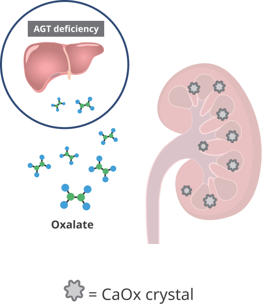
PH1 disease progression1,2
Excessive oxalate is produced in
the liver and combines with
calcium to form CaOx crystals.2

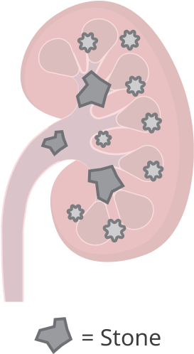
CaOx crystals aggregate to form
stones in the kidneys and can
also cause nephrocalcinosis.2

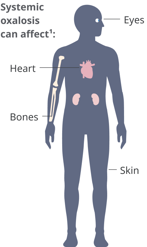
Disease progression often
leads to kidney failure and
systemic oxalosis.2
AGT=alanine-glyoxylate aminotransferase; CaOx=calcium oxalate; ESKD=end-stage kidney disease.
Early intervention in PH1 is critical and may delay renal deterioration5
PH1 progression is unpredictable; therefore, it should be treated early and monitored continuously.2,5 Most current standard-of-care approaches do not address the underlying cause of oxalate overproduction in PH1.2
Hyperhydration can be intense, uncomfortable, and difficult to maintain.2
Pyridoxine (vitamin B6) supplementation is only effective in reducing UOx levels in a subset of patients with PH1.2
Stone removal surgeries can cause unintended consequences.6 They often are only a temporary solution to a chronic problem and do not address the underlying cause of oxalate overproduction.2,3
Conventional dialysis often cannot remove enough oxalate to prevent PH1 disease progression, requiring increased treatment time and frequency.2
Dual liver-kidney transplant may eventually be required for patients with advanced CKD.2
CKD=chronic kidney disease; UOx=urinary oxalate
In a study
of patients with PH required a kidney stone removal surgery at least once, with many patients requiring multiple stone-removal procedures.7

PH1 can be challenging to diagnose8
Screening for primary hyperoxaluria should be undertaken if patients are presenting one or a combination of the symptoms listed below3,4,9-13:
- a single kidney stone in an infant or child <18 years old
- recurrent kidney stones in adults 18 years or older
- nephrocalcinosis
- family history of kidney stones
- elevated urinary oxalate (UOx) levels
- advanced CKD with unknown cause
- failure to thrive and ESKD in infants
- signs of systemic oxalosis
Analyzing specific genes is crucial for an accurate diagnosis in patients with kidney-stone diseases with clinical overlap in symptoms, as it helps identify disease-causing mutations. This can allow for patient management that may reduce recurrent symptoms or progression to ESKD.5,14,15
Important Safety Information for Rivfloza®
Adverse Reactions
- Most common adverse reaction (reported in 39% of patients) are injection site reactions, which include erythema, pain, bruising, and rash.
Indications and Usage
Rivfloza® (nedosiran) injection 80 mg, 128 mg, or 160 mg is indicated to lower urinary oxalate levels in children 9 years of age and older and adults with primary hyperoxaluria type 1 (PH1) and relatively preserved kidney function, eg, eGFR ≥30 mL/min/1.73 m2.
Please click here for Rivfloza® Prescribing Information.
References:
- Cochat P, Rumsby G. Primary Hyperoxaluria. N Engl J Med. 2013;369(7):649-658. doi:10.1056/NEJMra1301564
- Groothoff JW, Metry E, Deesker L, et al. Clinical practice recommendations for primary hyperoxaluria: an expert consensus statement from ERKNet and OxalEurope. Nat Rev Nephrol. 2023;19(3):194-211. doi:10.1038/s41581-022-00661-1
- Milliner DS, Harris PC, Sas DJ, Cogal AG, Lieske JC. Primary hyperoxaluria type 1. GeneReviews®. Updated February 10, 2022. Accessed October 2, 2023. https://www.ncbi.nlm.nih.gov/books/NBK1283/
- Hopp K, Cogal AG, Bergstralh EJ, et al; Rare Kidney Stone Consortium. Phenotype-genotype correlations and estimated carrier frequencies of primary hyperoxaluria. J Am Soc Nephrol. 2015;26(10):2559-2570. doi:10.1681/ASN.2014070698
- Fargue S, Harambat J, Gagnadoux MF, et al. Effect of conservative treatment on the renal outcome of children with primary hyperoxaluria type 1. Kidney Int. 2009;76(7):767-773. doi:10.1038/ki.2009.237
- Khan SR, Pearle MS, Robertson WG, et al. Kidney Stones. Nat Rev Dis Primers. 2016;2:16008. doi:10.1038/nrdp.2016.8
- Tang X, Bergstralh EJ, Mehta RA, Vrtiska TJ, Milliner DS, Lieske JC. Nephrocalcinosis is a risk factor for kidney failure in primary hyperoxaluria. Kidney Int. 2015;87(3):623-631. doi:10.1038/ki.2014.298
- Soliman NA, Nabhan MM, Abdelrahman SM, et al. Clinical spectrum of primary hyperoxaluria type 1: experience of a tertiary center. Nephrol Ther. 2017;13(3):176-182. doi:10.1016/j.nephro.2016.08.002
- Bhasin B, Ürekli HM, Atta MG. Primary and secondary hyperoxaluria: understanding the enigma. World J Nephrol. 2015;4(2):235-244. doi:10.5527/wjn.v4.i2.235
- Cochat P, Hulton SA, Acquaviva C, et al. Primary hyperoxaluria type 1: indications for screening and guidance for diagnosis and treatment. Nephrol Dial Transplant. 2012;27(5):1729-1736. doi:10.1093/ndt/gfs078
- Hoppe B, Beck BB, Milliner DS. The primary hyperoxalurias. Kidney Int. 2009;75(12):1264-1271. doi:10.1038/ki.2009.32
- Harambat J, Fargue S, Acquaviva C, et al. Genotype-phenotype correlation in primary hyperoxaluria type 1: the p.Gly170Arg AGXT mutation is associated with a better outcome. Kidney Int. 2010;77(5):443-449. doi:10.1038/ki.2009.435
- Frishberg Y, Rinat C, Shalata A, et al. Intra-familial clinical heterogeneity: absence of genotype-phenotype correlation in primary hyperoxaluria type 1 in Israel. Am J Nephrol. 2005;25(3):269-275. doi:10.1159/000086357
- Cogal AG, Arroyo J, Shah RJ, et al. Comprehensive genetic analysis reveals complexity of monogenic urinary stone disease. Kidney Int Rep. 2021;6(11):2862-2884. doi:10.1016/j.ekir.2021.08.033
- Braun DA, Lawson JA, Gee HY, et al. Prevalence of monogenic causes in pediatric patients with nephrolithiasis or nephrocalcinosis. Clin J Am Soc Nephrol. 2016;11(4):664-672. doi:10.2215/CJN.07540715
